Electrospun nanofibers when embedded within a transparent matrix is able to give a transparent composite. Moving beyond transparent composite, a stand-alone, thin layer of electrospun polymeric nanofibers mesh will also exhibit high level of optical transmittance. In air filtration, a thin layer of electrospun nanofibers mesh is able to capture micro-particles while showing good optical transparency. Using electrospun polyacrylonitrile nanofibers on a substrate, Liu et al (2015) demonstrated PM2.5 removal efficiency of more than 95% while allowing 90% transparency. Such a thin layer also reduces pressure drop when air passes through the membrane. High removal efficiency, transparency and low pressure drop makes the electrospun nanofibers membrane a potential window screen with filtration properties for indoor air protection.
Good optical transparency may also be achieved using highly aligned nanofibers. Kim et al (2016) constructed an electrospun membrane made out of randomly oriented nanofibers and some areas made out of highly aligned nanofibers. Sectors comprising of highly aligned nanofibers were created by having non-conductive cellophane tape on the rotating drum surface. The non-conductive cellophane tape creates a "gap" within a more conductive surface and this aligned fibers formed across the width of the cellophane tape similar in principle to the parallel electrodes collector for collecting aligned fibers. Comparison of the transparency using spectrum analysis showed that the aligned-oriented nanofibrous mat has a transparency that is twice higher than randomly oriented nanofibrous mat. However, it is unclear whether the greater transparency is due to fiber alignment or lower density of fibers in the aligned mat compared to the randomly oriented fiber mat.
Transparency due to fiber organization is not restricted to just parallel aligned fibers. Electrospinning may also be used to produce fibers that are radiating from a center. Kim et al (2018) used a non-conducting hemispherical device with a conducting pin in the middle and a wire that forms a ring at the base of the hemispherical device. This setup encourages the formation of a three-dimensional (3D) scaffold with electrospun fibers radiating from the center to the circular wire at the base. Due to the fiber organization, this scaffold was found to exhibit high transparency which is similar to native cornea in the visible wavelength when wetted in PBS.
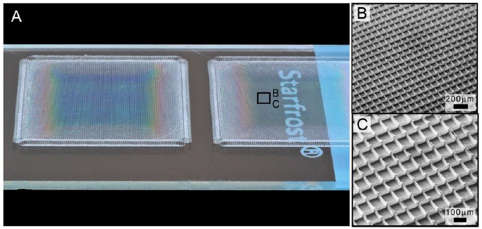
Progress in melt electrospinning and its combination with near-field electrospinning has led to the ability to fabricate ordered structure made of out fibers with diameter under 1 µm. Hochleitner et al (2015) was able to use melt electrospinning at close distance between the tip and the collector (1 to 10 mm) to produce polycaprolactone fibers with diameter of about 800 nm and stack them on top of one another to form an array of box-structures with periodicity of about 100 µm and height of 80 µm. Given the large area of empty spaces between the fibers, the underlying image can be seen clearly.
When constructing the membrane made from aligned fibers, the distance between the fibers need to be sufficiently large to maintain transparency. Pan et al (2015) was able to construct highly aligned silver nanowires (AgNWs) using near field electrospinning. To attain transmissivity of 70%, the nanofiber film thickness needs to be reduced to only 31 nm. At thickness of 292 nm, the transmissivity was reduced to only 15%. This is probably due to the density of the aligned nanofibers that reduces the amount of light that is able to pass through at higher thickness. Measured resistance of the AgNW film at 791 nm was 210.8 Ω/m but at such thickness, the film is likely to be opaque.
Light transmittance of electrospun fibers may be affected by the choice of polymer and not just the physical dimension. In an experiment by Chen et al (2015) using electrospun silk fibroin (SF), poly(l-lactic acid-co-ε-caprolactone) P(LLA-CL) and their mixture, the greatest transmittance is not due to fiber diameter or membrane thickness. Instead, it is from the 25:75, SF: P(LLA-CL) blend. Although the detail of why this is so was not investigated, it may be due to the molecular interaction between the two blended polymers. It may also be due to P(LLA-CL) nanofibers being more optically transparent compared to SF nanofibers.
Good transparency may also be obtained from electrospun fibers that swell upon exposure to fluid, forming a gel. Schulte-Werning et al (2021) was investigating the properties of electrospun fibers incorporated with several functionalities for potential use as wound dressing. The ingredients forming the blend were chloramphenicol (CAM), an antibiotic, water-soluble β-1.3/1.6 glucan (SBG®), an active ingredient in the topical treatment of diabetic foot and leg ulcers, chitosan (CHI), a bioactive polymer which exhibits intrinsic antimicrobial activity, hydroxypropylmethylcellulose (HPMC), a cellulose-based polymer with high swelling capacity, and polyethylene oxide (PEO) to facilitate fiber formation from electrospinning. The resultant electrospun fibers have an average diameter of 100 nm. Upon exposure to fluid, the multi-functional nanofiber membrane showed a high swelling index and became transparent. This helps to adhere to the wound and maintain a moist local environment. High transparency of the membrane also allows examination of the wound without the need to remove the dressing.
Several studies have also shown that sintered, inorganic nanofibrous membrane is optically transparent without a matrix material surrounding it. The resultant material is also flexible and may be used in a variety of applications from solar cell to flexible electronic screens.
Indium tin oxide (ITO) is a transparent conducting oxide that has been used in many applications. Electrospinning of its precursors, indium chloride tetrahydrate, tin chloride pentahydrate and poly(vinyl pyrrolidone) mixture has yielded nanofiber with diameter of about 220 nm. Although the resultant electrospun membrane is white, calcination at temperature above 400 °C forms pure ITO nanofiber membrane which is transparent. The transparent sheet is made of fibers with diameter of about 100 nm. Higher calcination temperature was shown to improve the optical transmittance through the membrane up to 92%. However, the membrane thickness needs to be very small with deposition duration as low as 20s to obtain the highest transmittance. When deposition duration was increased to 120s, the optical transmittance drops from 91% to 75% [Munir et al 2008]. While ITO on its own is transparent, other non-transparent inorganic material has been made transparent when the nanofibrous membrane is sufficiently thin.
Conductive and transparent film consisting of copper nanofiber network has been constructed on a glass substrate. Generally, precursor of copper is blended with a carrier polymers for electrospinning. Sintering was next carried out to remove the organic component and reduce the nanofiber to CuO. Lastly, the CuO nanofibers are reduced to form Cu nanofiber. Kim et al (2015) compared the optical transmission and electrical conductivity of randomly oriented copper nanofiber prepared using polyvinyl alcohol (PVA) and polyvinyl butyral (PVB) as carrier polymers for electrospinning of its precursor. Their studies showed that the optical transmittance was influenced by the amount of copper nanofibers and not the carrier polymers used but the electrical resistance were affected by the carrier polymers (See table 1). The poorer electrical conductivity of Cu nanofiber derived from PVA carrier was attributed to high reduction temperature used compared to PVB and this led to discontinuity of the nanofibers on the substrate.
Wu et al (2010) used copper acetate/polyvinyl acetate solution for electrospinning to give nanofibers. With a thin nanofiber layer, the optical transmittance is excellent in the visible and near-infrared ranges. Aligned Cu nanofibers was able to show 90% transmittance at sheet resistance of 25 ohm/sq[Wu et al 2010]. Transparent and flexible electrodes have been constructed by transferring the Cu nanofiber network to poly(dimethylsiloxane) (PDMS) substrate [Wu et al 2010].
To improve the thermal and chemical resistance of copper nanofiber membrane, Hsu et al (2012) used atomic layer deposition to coat a passivation layer of aluminum-doped zinc oxide (AZO) and aluminum oxide on copper nanofibers. Deterioration in the conductivity of the copper membrane is investigated by measuring its resistance. Unprotected copper nanofibers quickly become insulating when it undergo thermal oxidation at 160 °C in dry air and 80 °C in humid air (80% relative humidity). However, Cu nanofiber with AZO/Al
2O
3 layer showed an increased sheet resistance of only 10% after baking the fibers at 160 °C for 8 h. When bare copper nanofibers were coated and baked with an acidic layer of PEDOT:PSS, its sheet resistance increased by 6 order of magnitude while protected copper nanofiber showed an 18% increase demonstrating the superior protection offered by AZO/Al
2O
3 layer. With the coating, optical transmittance over the visible light wavelength range showed less than 1% decrease with no effect on the electrical conductance of the Cu nanofibers.
An alternative method of fabricating metal nanowire on a transparent substrate using electrospun nanofibers is to load reducing agent in the fiber followed by immersion in a salt solution for reduction of the ions form a continuous coating over the nanofiber. This method is demonstrated by Hsu et al (2014) to form Ag or Cu nanowire. In their process, they electrospun polyvinyl butyral (PVB) loaded with tin (II) chloride (SnCl2) fibers onto a glass substrate. The PVB/ SnCl2 fibers were immersed in silver nitrate solution for reduction of Ag+ to Ag. At 70 nm metal deposition thickness, the metallization coverage and optical transmittance was good. Resistance and transmittance for Ag nanowire was 8.5 Ω/sq and 90% respectively.
Stretching the concept of using electrospun nanofibers in the construction of conductive linkages, conductive nanotroughs have been fabricated using electrospun nanofibers as a template material. A variety of methods are available for coating a conductive layer on electrospun nanofibers. Metallic coatings such as chromium, gold, copper, silver and aluminum can be applied to electrospun nanofibers using thermal evaporation [Wu et al 2013]. Platinum and nickel has been coated on electrospun nanofibers using electron-beam evaporation and silicon and ITO using a.c. magnetosputtering [Wu et al 2013]. The coated nanofiber sheet can be transferred to a silicon substrate using dropcast. With a coating thickness of 100 nm on one side, the nanotrough layer is sufficiently strong to be self-supporting after the nanofiber templates have been removed [Wu et al 2013]. A single gold nanotrough was found to have an electric conductivity of 2.2 x 10⁵ S/cm which is slightly less than its polycrystalline bulk [Wu et al 2013]. The nanotrough network exhibit greater transparency than flat nanostrips due to its concave shape which reduces its electromagnetic cross-section with transmittance of more than 90% for Cu and Au nanotrough materials across the visible wavelengths. It is also highly bendable, stretchable and foldable without significant deterioration in electrical conductivity [Wu et al 2013].
Published date: 29 Jan 2014
Last updated: 20 September 2022
▼ Reference
-
Chen J, Yan C, Zhu M, Yao Q, Shao C, Lu W, Wang J, Mo X, Gu P, Fu Y, Fan X.
Electrospun nanofibrous SF/P(LLA-CL) membrane: a potential substratum for endothelial keratoplasty. International Journal of Nanomedicine 2015; 10: 3337.
Open Access
-
Hochleitner G, Jungst T, Brown T D, Hahn K, Moseke C, Jakob F, Dalton P D, Groll J. Additive manufacturing of scaffolds with sub-micron filaments via melt electrospinning writing. Biofabrication 2015; 7: 035002.
Open Access
-
Hsu P C, Wu H, Carney T J, McDowell M T, Yang Y, Garnett E C, Li M, Hu L, Cui Y. Passivation Coating on Electrospun Copper Nanofibers for Stable Transparent Electrodes. ACS Nano 2012; 6: 5150. doi: 10.1021/nn300844g
-
Hsu P C, Kong D, Wang S, Wang H, Welch A J, Cui Y. Electrolessly Deposited Electrospun Metal Nanowire Transparent Electrodes. J. Am. Chem. Soc. 2014; 136: 10593.
-
Kim S, Lee H, Kim D, Ko D, Kim D, Kim H, Yoon S G. Transparent Conductive Films of Copper Nanofiber Network Fabricated by Electrospinning. Journal of Nanomaterials 2015; 2015: 518589.
Open Access
-
Kim J I, Hwang T I, Aguilar L E, Park C H, Kim C S. A Controlled Design of Aligned and Random Nanofibers for 3D Bi-functionalized Nerve Conduits Fabricated via a Novel Electrospinning Set-up. Scientific Reports 2016; 6: 23761.
Open Access
-
Kim J I, Kim J Y, Park C H. Fabrication of transparent hemispherical 3D nanofibrous scaffolds with radially aligned patterns via a novel electrospinning method. Scientific Reports 2018; 8: 3424.
Open Access
-
Liu C, Hsu P C, Lee H W, Ye M, Zheng G, Liu N, Li W, Cui Y. Transparent air filter for high-efficiency PM2.5 capture. Nature Communications 2015; 6: 6205.
-
Munir M M, Widiyandari H, Iskandar F, Okuyama K. Patterned indium tin oxide nanofiber films and their electrical and optical performance. Nanotechnology 2008; 19: 375601.
-
Pan C T, Yang T L, Chen Y C, Su C Y, Ju S P, Hung K H, Wu I C, Hsieh C C, Shen S C. Fibers and Conductive Films Using Silver Nanoparticles and Nanowires by Near-Field Electrospinning Process. Journal of Nanomaterials 2015; 2015: 494052
Open Access
-
Pan C,
Wu H, Hu L, Rowell M W, Kong D, Cha J J, McDonough J R, Zhu J, Yang Y, McGehee M D, Cui Y. Electrospun Metal Nanofiber Webs as High-Performance Transparent Electrode. Nano Lett. 2010; 10: 4242.
-
Schulte-Werning LV, Murugaiah A, Singh B, Johannessen M, Engstad RE, Skalko-Basnet N, Holsæter AM. Multifunctional Nanofibrous Dressing with Antimicrobial and Anti-Inflammatory Properties Prepared by Needle-Free Electrospinning. Pharmaceutics. 2021; 13(9):1527.
Open Access
-
Wu H, Kong D, Ruan Z, Hsu P C, Wang S, Yu Z. A transparent electrode based on a metal nanotrough network. Nature Nanotechnology 2013; 8: 421.
▲ Close list
 ElectrospinTech


