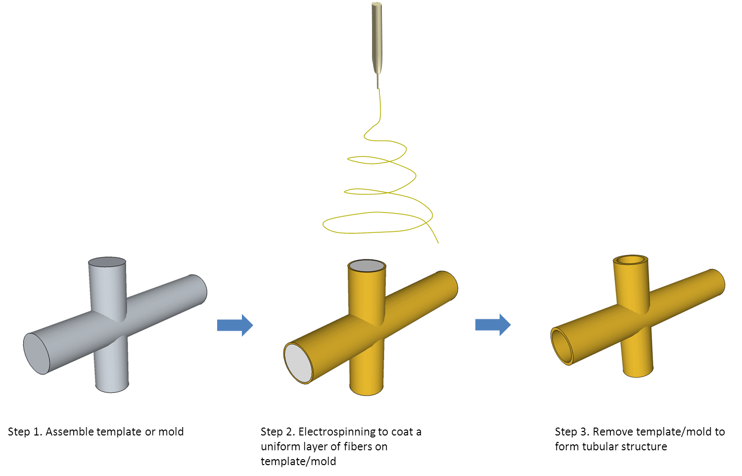
The electrospinning process typically "sprays" a layer of fibers which can be deposited or coated on substrates. Given a template and sufficient deposition time, a fibrous structure taking the form of the underlying substrate may be constructed. The most common structure fabricated through this method is small diameter tubes where fibers are coated on a rotating rod and thereafter, the template rod is removed. Using the same concept, three-dimensional nanofibrous tubes with junctions have been constructed [Zhang et al 2008]. First, rod templates forming the desired configuration are assembled. Electrospinning is used to coat the full surface of the template until sufficient thickness is achieved. Finally, the internal rods are removed thus forming free standing tubular fibrous structure.
Electrospun scaffold have been demonstrated in tissue cultures to facilitate cell adhesion and proliferation. However, for actual tissue or organ replacement, the shape and structure of the scaffold needs to be tailored to the specific application. Electrospinning may be used in combination with other manufacturing techniques to construct customized structures. With development of 3D scanning and imaging techniques, rapid prototyping can be used replicate the shape of the organs or tissues that require replacement. Adjustment to the 3D CAD model may be made prior to the construction of the template mold using rapid prototyping. The final physical model may then be used as a substrate for layering of electrospun nanofibers to form the final product. This method has been demonstrated by Owida et al (2011) to fabricate structurally optimized coronary artery bypass graft.
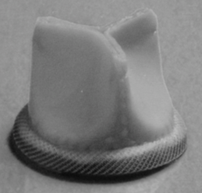
Other more complex structure has also been fabricated using the template coating method. Electrospun trileaflet heart valve prosthesis has been constructed [Gaudio et al 2007] by depositing a uniform coating of nanofibers on a trileaflet heart valve-shape metallic template through slow rotation of the template (0.3 to 0.4 rpm). In vitro functional testing of the leaflets showed satisfactory coaptation in the diastolic phase and synchronous opening thus demonstrating the technical feasibility of using this method for the construction of the heart valve.
Creating a nanofibrous structure using a template is not limited to biomedical applications. Creative use of this concept has led a start-up company, Electroloom, to design a machine for making garments which can be peeled off straight from the template and ready to be worn after electrospun fiber deposition.
In general, templates or molds without undercut are easier to fabricate. Narrow gaps, cavities, concave surfaces, openings and uneven surfaces should also be avoided since fibers may deposit across higher grounds and not reaching the inner surface. A greater margin of tolerance for the uniformity of the fiber thickness is required for uneven surfaces since fibers tend to deposit preferentially on higher or protruding surfaces.
For electrospinning on a template, a challenge is on getting the fibers to deposit on any recess or depression on the template. To replicate organs that have undulating surfaces, this limitation of electrospinning needs to be addressed. Song et al (2021) demonstrated the feasibility of using a hydrogel collector to construct a 3D ear using electrospinning. An advantage of hydrogel is that it is flexible and can be flattened to give a more uniform surface for fiber deposition. Hydrogel would also contain mobile ions in the interstitial fluid such that it can attract the incoming electrospinning jet as well as a metal collector. The hydrogel may be restored to its original shape after a layer of fibers have been collected. Song et al (2021) selected a mixture of alginate and gelatin as their hydrogel template. Flattening of the alginate/gelatin hydrogel collector enables fiber to be deposited within the complex geometries of helix and antihelix of the ear. The thickness of the deposited fibers across the surface of the flattened hydrogel ear template was found to be uniform in contrast to the unflattened, original 3D ear shaped hydrogel collector where no fibers were deposited in the indented part. Although this method is able to get fibers to be deposited within the indentation of a structure, it may not be suitable if the intention is to create a replica of the structure consisting of electrospun fibers only.
Published date: 02 Jan 2014
Last updated: 12 October 2021
▼ Reference
-
Del Gaudio C, Bianco A, Grigioni M. Electrospun bioresorbable trileaflet heart valve prosthesis for tissue engineering: in vitro functional assessment of a pulmonary cardiac valve design. Ann Ist Super Sanita 2007; 44: 178.
-
Owida A, Chen R, Patel S, Morsi Y, Mo X. Artery vessel fabrication using the combined fused deposition modeling and electrospinning techniques. Rapid Prototyping Journal 2011; 17: 37.
-
Song J Y, Ryu H I, Lee J M, Bae S H, Lee J W, Yi C C, Park S M. Conformal Fabrication of an Electrospun Nanofiber Mat on a 3D Ear Cartilage-Shaped Hydrogel Collector Based on Hydrogel-Assisted Electrospinning. Nanoscale Res Lett. 2021 Jul 9;16(1):116.
Open Access
-
Zhang D, Chang J. Electrospinning of Three-Dimensional Nanofibrous Tubes with Controllable Architectures. Nano Letters 2008; 8: 3283.
▲ Close list
 ElectrospinTech

