Home | About | Contact


Stacking layers of fibers

LBL stack 3D nanofiber
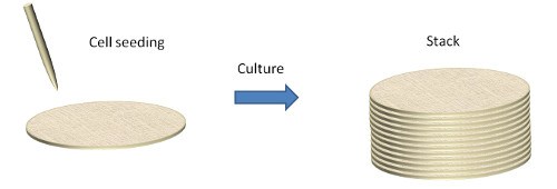
Layer by layer stacked 3D nanofiber schematic

Generally, the typical output of a standard electrospinning setup is a two-dimensional compact membrane. As the deposition duration increases, the membrane will grow in thickness but the build-up is slow. In tissue engineering, a thicker scaffold is often necessary to fill up a volume defect. Stacking fiber membranes is a much faster way of achieving sufficient volume thickness due to the presence of interface between layers. Another advantage of this method is that it allows the fabrication of a thick scaffold composed of aligned fibers in a single direction. Orr et al (2015) used a pair of ceramic magnet - copper electrode set and arranged in parallel at 10 cm apart over a water bath to collect aligned nanofibers. The water bath below the parallel electrodes allows the collected fibers to rest on the surface of the water at its center. The aligned fibers can be easily lifted off the water and the parallel electrodes and stacked together.

Electrospun membranes stacked on one another needs to remain together as a whole structure. One method of maintaining the integrity of the membrane-stacked structure is to have cells that are first cultured on the individual membrane before putting them together for a few more days to fuse the layers. To demonstrate the viability of culturing cells on separate layers and stacking, He et al (2015) used this technique to construct a cell-based 3D cartilage scaffold. First, they place a single sheet of gelatin/polycaprolactone nanofibrous membranes in a well of 6-well plate. A cell suspension of bone marrow stromal cells and chondrocytes were seeded on the nanofibrous membrane. Another sheet of nanofibrous membrane was stacked on top of the first sheet followed by cell seeding. This was repeated until there were ten sheets of nanofibrous membranes. The cell-scaffold construct was cultured in vitro for 1 week before implantation into nude mice. Another advantage of this technique is that the complex distribution of cell types in an organ can be replicated. The distribution of cells in the skin is such that the inner layer comprises mainly of fibroblast while the upper layer closer to the skin surface is mainly populated by keratinocytes. With this organization in mind, Yang et al (2009) cultured fibroblast and keratinocytes on separate electrospun PCL/collagen membranes. These membranes were constructed by alternating electrospinning of fibers directly into a cell culture media and seeding of cells. The layers were built up such that the fibroblast layers were at the bottom and the keratinocyte layers above. After culturing for 3 days, the individual layers were found to be tightly bounded together. Electrospinning and deposition of hydrogel may be carried out alternately to form a 3D layered construct. Xu et al (2013) used a hybrid printing device for alternate electrospinning polycaprolactone (PCL) and inkjet printing of rabbit elastic chondrocytes suspended in a fibrin-collagen hydrogel. This creates a one-step method of constructing layered scaffolds with cells seeded without the need for manual membrane transfer and cell seeding.

Binding agents may also be used to bond the layers together or to encapsulate the layers and this has been demonstrated through the use of hydrogel [Yang et al 2011]. Introducing a second solution after the fiber layers have been stacked up followed by thermally induced phase separation of the second solution have also been used to bind the layers[Vaquette et al 2013]. This method has the added advantage of introducing porous interfaces between the fibrous layers. Heat treatment with pressurized gas has also been shown to be effective in fusing the fiber layers. The resultant 3D scaffold showed much better modulus and strength compared to untreated scaffold [Leung et al 2012]. Cold welding under high pressure has also been used to create fusion between the fiber-layers However, this method was shown to significantly reduce the porosity and air permeability through it [Madurantakam et al 2013]. In clinical application, fibrin gel may be used as the binding agent. Beachley et al (2014) seeded myoblast of several sheets of polycaprolactone before stacking them together and using fibrin gel between the layers for adhesion and encapsulation of the cells. Yang et al (2019) used gelatin to bind layers of electrospun polycaprolactone (PCL) together. Individual layers of PCL mesh was first dipped in gelatin solution. There after, the gelatin coated PCL meshes were stacked together and cross-linked in 1% genipin solution for 24hrs. This way they were able to construct scaffolds made of aligned nanofiber mesh and randomly oriented nanofiber mesh. Gelatin has better cell adhesion and biocompatibility than PCL and cultured human periodontal ligament mesenchyme cells (PDLSCs) were found in greater numbers in the gelatin film between the PCL layers. Since gelatin biodegrade at a faster rate than PCL, the adhered cells may be transferred to the PCL surface and their secreted ECM may take over the binding of the PCL layers to maintain the structural integrity of the 3D scaffold.

Another method to combine the layers is to use sintering. Wright et al (2010) used vapour and heat sintering to fuse layers of poly(d,l-lactide) and poly(l-lactide) electrospun fibers. For vapour sintering, the layers were fused by exposure to tetrahydrofuran vapour (240 °C) for 10 minutes while heat sintering was carried out at 54 °C for 30 minutes. An advantage of sintering is that it increases the mechanical strength of the material due to fusion between the fibers. Between the two sintering process, heat sintering is preferred as vapour sintering requires more accurate control. Slight extension in the duration of vapour sintering condition has been shown to cause significant degradation in the fibrous structure.


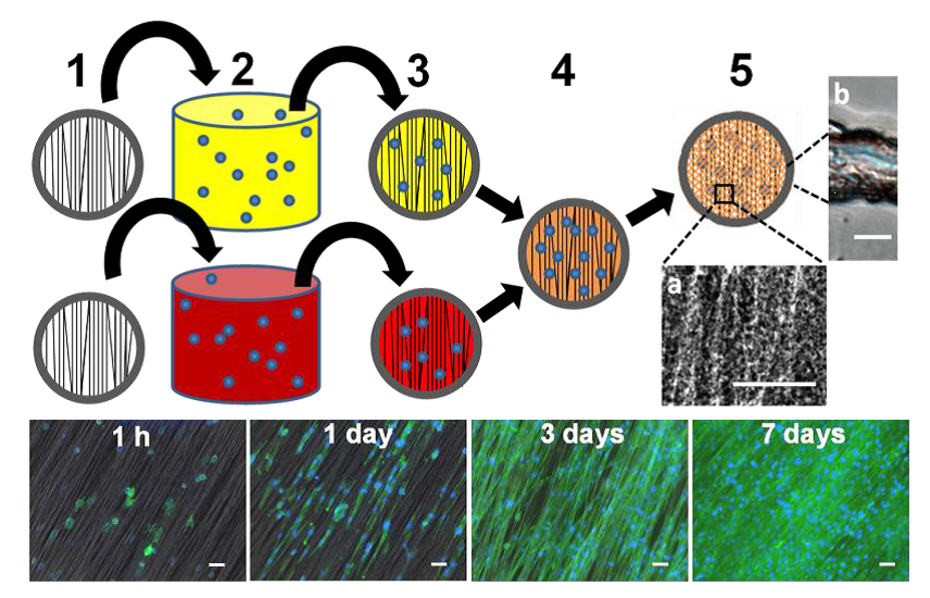
(Top) One step direct cell seeding with fibrin gel. (1) Mounted nanofiber arrays are immersed in (2) fibrinogen (represented in yellow color) and thrombin (represented in red color) solutions containing suspended cells (represented in blue color), respectively. (3) Removal from the liquid results in the formation of a thin, uniform liquid film over the nanofiber arrays. (4) Fibrinogen and thrombin cell-containing films are sandwiched together and in close contact. (5) A polymerization reaction results in crosslinked fibrin film with embedded nanofibers and cells. (a) A close up image of fibrin filaments surrounding the aligned polycaprolactone (PCL) fibers (scale bar = 50 µm). (b) a cross-section of a composite fibrin gel nanofiber composite film (scale bar = 10 µm). (Bottom) Fluorescent images of nanofiber/cell sheets with direct C2C12 cell fibrin seeding taken at 1 h and 1, 3 and 7 days after fabrication. Actin filaments were stained in green, and cell nuclei were stained in blue. Scale bar = 50 µm. [Beachley et al. Bioengineering 2014; 1: 114. This work is licensed under a Creative Commons Attribution 4.0 International.]

Electrospinning may be used to create alternate layers of a desired polymer material and a low melting point polymer material. Rashid et al (2020) constructed a multi-layered scaffold made of mainly electrospun polydioxanone (PDO) aligned fibers, electrospun polycaprolactone (PCL) random fibers and woven PDO fibers. PCL with its lower melting point was used as a bonding agent to hold the scaffold together. A hot compressed heat treatment of 80 °C for 1 - 3 min was used to melt the PCL mesh which has a melting temperature of 65 °C. This creates a biodegradable scaffold with the faster degrading PDO being held together by the slower degrading PCL electrospun fibers.

Advantages for tissue engineering :
  • Even distribution of cells on each membrane and throughout the stacked 3D structure
  • Easy isolation of infected population prior to putting the membranes together
  • Standard cell culture technique
  • Each membrane may have its own distinctive properties.
Disadvantages
  • Little or no migration of cells through the layers.
  • Distinct interface between layers.
  • Difficult to scale up

The ease of removing the nanofibrous membrane from the collector surface varies depending on the polymer used. Typically, a synthetic polymer is easier to peel off compared to natural polymers such as collagen. There are a few broad concepts that can be used to facilitate seprarating of the nanofibrous membrane from the collector surface. One, a separating medium (eg. dental or medical grade alginate solution) may be coated on the collector surface prior to fiber deposition. Two, use a collector with a non-stick coating (Teflon) on its surface. Three, use a non-conventional collector such as depositing the fibers on water [Tzezana et al 2008]. If the membrane size is small, a ring or cylinder(with height of 5 mm and above) may be used. In the later method, the electrospinning jet will coat the open end of the cylinder with nanofibers. The membrane covering the end of the cylinder may be removed by pushing a rod against it.

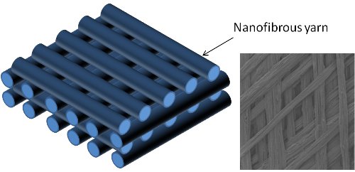
Without modifications to the setup, electrospun nanofibrous membrane are known to present a barrier to cell penetration [Pham et al 2006]. Cell seeded on the top surface of a 3D scaffold constructed from stacked membranes will have difficulty migrating to the inner layers. To overcome this limitation of stacked membranes, strips of nanofiber membrane or nanofiber yarns may be used as beams to form layered grids. Heat treatment may be used to fuse the nanofibrous strips [Cai et al 2012]. The distance between each nanofiber strip can be controlled to give the desirable pore size. One way to mechanize the layering process is to use a roller setup with a continuous nanofibrous yarn fabrication process.

While stacking of the membranes can be carried out after it has been peeled off from the collector surface, another way is to stack the membranes while it is still on the collector. For this, thin aluminium sheets with multiple matching holes may be used to collect the fibers. If the hole is relatively small, less than 2 cm diameter, it can be easily covered by the deposited fibers. Another piece of aluminium sheet with holes may be positioned over it such that the position of the holes matches. A fresh layer of electrospun fibers may be collected on the aluminium sheet. After several layers of aluminium sheets have been stacked, the aligned layers of electrospun fibers may be removed by using a punch to cut through the holes covered by the fibers. An advantage of this method is that the pore sizes between fibers covering the holes are larger than the fibers collected on a solid surface [Hong et al 2015].

Layered stacked yarns
Using nanofibrous yarns or taps to form layered grids.
Concentric fiber layer

A variation of the stacked membrane layer is to roll a piece of membrane to form a cylindrical scaffold. To prevent the clyindrical scaffold from "unrolling", binders (eg. fibrin glue) or heat treatment [Leung et al 2012] may be used to fuse the fibers. This structure is relatively easy to construct and is less tedious than stacking layers of membrane. For applications where the presence of a small gap or interface along the length of the scaffold is preferred (eg. cell migration across a gap), this arrangement of the membrane will be useful. Deng et al used this arrangement to replicate cortical bone structure and the seeded osteoblast was able to bridge the gap between the concentric walls [Deng et al 2011]. Wright et al (2010) created concentric roll of poly(d,l-lactide) and poly(l-lactide) electrospun fibers strip and used sintering to fuse the structure. Depending on the application of rolled concentric nanofibrous layers, bonding agents may not be needed if it is held within another structure. Yun et al (2019) electrospun a poly(lactide-co-glycolide)/hydroxyapatite nanofibres sheet and roll into a thick wall cylinder with wall thickness of 2 mm. A 3D melt printing method was used to replicate the radius bone segment of the study subject, beagle dog. The 3D printed poly(lactic-acid) scaffold has a hollow in the center which was filled in with the rolled poly(lactide-co-glycolide)/hydroxyapatite nanofibres prior to implantation into the segmental bone defect. The rigid 3D printed poly(lactic-acid) scaffold was able to prevent the rolled poly(lactide-co-glycolide)/hydroxyapatite nanofibres from unraveling.

Cylindrical roll
Manual rolling of membrane to form a concentric layer of fibers.

Small pore size of electrospun membrane is known to restrict cell penetration into its depth. Although cells are able to migrate along the layers of the concentric 3D scaffold, they are unlikely to migrate through the layers. Joshi et al (2013) overcome this limitation by first using lasers to create macropores on the flat membrane before rolling onto a catheter. The patterns are created such that there will be overlapping between the pores when it is rolled. The shape of the rolled scaffold is maintained using silicone plug such that any cell migration will be through the surface of the scaffold instead of from the ends. Using scaffolds with pore sizes of 80 µm, 160 µm and 300 µm and implanted in the peritoneal cavity of Lewis rats after wrapping in omentum, they found that only scaffold with 300 µm exhibits full cell penetration after two weeks and significant vascular ingrowth to a distance of 850 µm. This shows that scaffold with sufficiently large pore size is required for host tissue integration. Wright et al (2010) used salt particles leaching to create pores within the concentric fibrous roll. However, in their study, the pore size created by the salt particles were not measured. To create space between the walls of a rolled up electrospun membrane, Kosowska et al (2020) coat the membrane with a gelling agent prior to rolling into a cylinder. The construct was frozen before immersing in a gelling solution to form the final scaffold. The steps of coating the membrane and freezing the construct is important to maintain a gap between the overlapping membrane. The distance between walls in the final scaffold was found to range from 50 to 200 µm. Kosowska et al (2020) used polylactide/hydroxyapatite (PLA/HA) as the electrospun fiber membrane and chitosan/graphene (CS/GO) as the hydrogel in the scaffold. Degradation of chitosan would further expose the gap between the walls for cell infiltration.


Published date: 23 August 2012
Last updated: 15 February 2021

Comments
[+]


Google Ad.