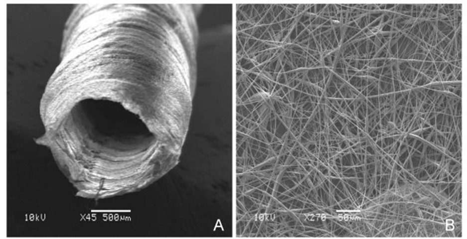
Numerous studies have been carried out to investigate the potential use of electrospun scaffold in peripheral nerve regeneration. One of the earliest electrospun scaffolds used in in vivo study of peripheral nerve regeneration in a sciatic nerve rat model is a simple conduit made out of randomly oriented nanofibers (Material: poly(L-lactide-co-glycolide)) [Bini et al 2004]. The results showed positive reflex responses in 45% of the rats with the implant and a thin fibrous tissue capsule on the exterior of the conduit after a month. A similar study by Panseri et al (2008) using randomly oriented polycaprolactone and poly(DL-lactide-co-glycolide) also demonstrated potential use of electrospun conduit as peripheral nerve regeneration graft. However, Duda et al (2014) reported strong foreign body reaction when electropun polycaprolactone fibers were used as a sheath material in their rat sciatic nerve model. Although it is not apparent what is the reason for the poor result since polycaprolactone is known for its biocompatibility, a possible reason could be the larger diameter of their fibers with average diameter of about 7 µm. Larger diameter fibers are generally less biocompatible than smaller fiber diameter scaffold (Read more) . Yu et al (2011) showed that with collagen/poly(e-caprolactone) (1:1) randomly nanofibrous (fiber diameter of 675 nm) conduit, no apparent neuroma formation or serious chronic inflammatory reaction was observed at 4 months. Other peripheral nerve grafts are more advanced in terms of fiber organization, presence of intra-luminal guidance channel and biomolecules incorporation.
The main purpose of the peripheral nerve graft is to facilitate axon neurite extension to bridge the gap and to prevent cell migration either into or out of the graft. In a study where perforations (pore size of 200 µm) were made on an electrospun nonwoven chitosan nerve conduit, granulation tissues and accompanying blood vessels were found to invade the lumen of the conduit [Rodotheoou et al 2013]. Not surprisingly, test results on the recovery of the peripheral nerve were similar or worse than empty conduit although the perforations did increases the number of blood vessels within the defect site. In vitro studies have shown that aligned fibers are able to guide neurite growth and this feature has been used in peripheral nerve graft for in vivo studies [Wang et al 2008]. The benefit of having longitudinally aligned fibers in the conduit has been found in several in vivo rat sciatic nerve studies. A comparison of conduits made out of randomly oriented chitosan nanofibers and longitudinally aligned chitosan nanofibers showed better results with longitudinally aligned nanofibrous conduit in electrophysiological tests, axon myelination and orientation and von Frey Hair Test [Wang et al 2008]. Zhu et al (2011) did similar comparison with poly (L-lactide-co-caprolactone)/poly(propylene glycol) nanofibrous conduit. Electrophysiological tests showed better performance of luminal aligned fibers conduit at 2 months compared to autograft and randomly aligned fibers conduit and no significant difference between luminal aligned fibers conduit and autograft at 12 months while randomly aligned fibers conduit continued to fare poorly. Fibrous thickness layer in the lumen of randomly aligned fibers was also found to be much thicker than the layer found in aligned fibers at 12 months. Myelin sheath thickness in aligned fibers was thicker in aligned fibers than randomly aligned fibers at 12 months but autograft showed highest density of thicker myelin sheath. When poly(e-caprolactone-co-ethyl ethylene phosphate conduits with circumferentially aligned fibers was compared to conduits with longitudinally aligned fibers, rat implanted with the later conduit also demonstrated better recovery [Chew et al 2007].
A potential drawback of hollow fibrous conduit is the possibility of conduit collapse which can be as high as 40% to 50% of the cases [Panseri et al 2008; Koh 2009]. A collapsed conduit will prevent migration of axon across the gap. Further, for larger gap, projection of the axon growth cone will be non-directed. Random movement of the growth cone will lengthen the journey and time taken to re-establish link with the distal endoneurial tube. The function of a guided nerve tube is to provide a path for the axon to follow across the gap. Studies of electrospun conduit with longitudinally aligned inner wall surface has already demonstrated better treatment outcome in in vivo rat models. Therefore, structures with longitudinally aligned nanofibrous fibers placed in the lumen of the conduit may further accelerate recovery.
A significant advantage of conduit with intraluminal guidance structures over empty conduit is the maintenance of conduit integrity whether the structure is made out of films [Clements et al 2009], yarns or monofilaments [Jha et al 2011]. However, while these structures guide axon extension, they are also a source of obstruction and this may affect recovery of the injured site. Clements et al (2009) compared the recovery of rat sciatic nerve injury using conduit with one and three luminal guidance sheets. Conduit with a single luminal guidance sheet showed better results in most tests compared to conduit with three guidance sheets. The exception is the area of regeneration cable which is found in greater number for three guidance sheets thus providing evidence that the guidance sheet does facilitates cable regeneration. A limitation of using sheets as guidance structure is that it invariably creates compartments within the lumen. Cells at the empty regions within each compartment are not guided by the sheet and may result in random organization as shown by misaligned fibroblasts, poorly aligned axons, Schwann cells and disorganized collagen bands. For more uniform distribution of guidance surface across the conduit cross-section for axon and Schwann cells migration, Koh (2009) used nanofibrous yarn as guidance structure. Each yarn is made out of nanofibers that are aligned along its length and a bundle of yarns are inserted in the lumen of a nanofibrous conduit. The inner surface of the conduit was made out of longitudinally aligned nanofibers while the outer surface was made out of randomly aligned nanofibers. Comparison of tests such as sensory recovery test, muscle mass ratio, electrophysiological, axon density and diameter showed similar or better results for conduit with the guidance structure to those without. However, neurophysiological, axon density and diameter were better for autologous graft while conduit with intraluminal yarns showed similar results to autologous graft for sensory recovery test and muscle ratio test. For the best balance between guidance distribution and minimal structural obstruction to axon and cell migration, Jha et al (2011) used nanofibrous monofilaments aligned along the length of conduit within its lumen. Their preliminary results showed 25% of axons present in the proximal section reached the distal end at 7 week interval. Unfortunately, no other tests were performed.
Zennifer et al (2025) used 3D printing to fabricate thermoplastic polyurethane (TPU) fiber lattice and electrospinning to deposit poly(3-hydroxybutyrate-co-3-hydroxyvalerate) (PHBV) fibers on it. The bilayered mesh was subsequently rolled into a spiral and heat sealed at the edges to form TPU/PHBV nanofibers guided conduits (NGCs).
In vivo tests using 10 mm sciatic nerve defect in Wistar rats showed good muscle innervation, axon healing and functional recovery comparable to autografts over 4 months. Sciatic functional index (SFI), toe-out angle and myelinated area were similar for both autograft and TPU/PHBV NGCs. Significant gastrocnemius muscle reduction was seen in both autograft, and TPU/PHBV implanted groups with gastrocnemius muscle index (GMI) of 54.22, and 43.67, respectively while the positive control (normal animal) was 92.04.
To reduce obstruction by guidance structure, another method is to tailor the degradation rate of the material such that the resorption of the guidance channel matches that of axon growth.
Physical structure of the conduit has been found to play an important role in promoting functional recovery in peripheral nerve injury. Another important consideration is the use of biomolecule to facilitate recovery. Bioactive compounds may be introduced to the electrospun scaffold using covalent bonding, blending, direct injection or core-shell nanofibers. Wang et al (2008) covalently bonded amino acids CYIGSR, CGGYIGSR (CG2YIGSR), or CGGGGGGYIGSR (CG6YIGSR) to chitosan nanofibrous conduit inner surface and showed similar or slightly better results than chitosan conduit without treatment. Chew et al (2007) showed that conduits made out of longitudinally aligned fibers with human glial cell-derived neurotrophic factor (GDNF) blended was able to show better functional recovery compared to conduits without. Koh (2009) used both biochemical cues and physical cues in her conduit construct. The physical cues are luminal guidance yarn and chemical cues are blended nerve growth factor and laminin. Between laminin and nerve growth factor, groups with laminin seem to perform slightly better than those without.
Increased local exudation and inflammatory reactions in peripheral nerve injury may have a negative impact on nerve regeneration and repair. Therefore, inhibiting overactive inflammatory response at the injury site may facilitate peripheral nerve repair. Xu et al (2022) loaded electrospun poly (lactic-co-glycolic acid) (PLGA) nanofibrous scaffold with Tacrolimus (FK506), an FDA-approved immunosuppressant, to improve treatment outcome. The electrospun PLGA/FK506 membrane was rolled into a conduit and tested in a rat sciatic nerve model with a 15 mm nerve segment resected. PLGA scaffold without FK506 loaded was used as a negative control. After 12 weeks, gait analysis, electrophysiology, and neuromuscular histology results showed that PLGA only scaffold performed significantly worse than PLGA/FK506 scaffold. There was no significant difference in the performance between PLGA/FK506 scaffold and autologous graft. The addition of FK506 into electrospun PLGA scaffold is a promising method for repairing peripheral nerve injury.
Nanofibers with core-shell structure containing bioactive compound in the core may be used to slow the compound release compared to a blended system where the compound is distributed throughout the fiber. Liu et al (2011) constructed core-shell poly(lactic acid-caprolactone) nanofiber with nerve growth factor and bovine serum albumin in the core for implantation in rat sciatic nerve model. Although the conduit was made of randomly oriented nanofibers, they reported recovery at twelve weeks that is comparable to autograft and much better than empty conduit and conduit with nerve growth factors injected in terms of regenerated myelinated nerve fibers number, axon diameter, myelin thickness, gastrocnemius muscle weight ratio, sciatic functional index and electrophysiological study.
Nerve injury recovery has been known to benefit from electrical stimulation. Wang et al (2010) seek to incorporate electrical stimulation in electrospun graft by constructing a chitosan nanofiber (randomly oriented) mesh conduit containing electrically polarized b-tricalcium phosphate particles. Examination of the electrically polarized composite graft at 12 weeks post implantation in a rat sciatic nerve model showed no significant difference to autograft in electrophysiological tests and histological tests.
Biodegradable latic- or glycolic- based polymers are often used in the construction of nerve conduits. However, their degradation may increase the acidity of the surrounding environment which is detrimental for recovery. To mitigate this issue, Lin et al (2017) loaded electrospun poly(D,L-lactic acid) (PDLLA) fibers with β-tricalcium phosphate (β-TCP) to neutralize the acidic residues from the degradation of PDLLA. Collagen was also added to the composite to improve biocompatibility. In their in vivo study using Wister rat sciatic nerve model, with electrospun conduit made of PDLLA, PDLLA/β-TCP, collagen/β/PDLLA and autologous graft, they found that nerve regeneration of collagen/β/PDLLA group was close to that of autologous graft, both of which were better than PDLLA and PDLLA/β-TCP groups after 3 months. While the presence of collagen may seems to have a more significant impact on regeneration, more studies would be needed to verify the benefits of having β-TCP.
The gold standard for peripheral nerve repair is the use of isograft or autograft and a key reason for its efficacy is the presence of Schwann cell. With advances in tissue engineering, cell seeded peripheral nerve repair graft may be used to replicate the advantages of autograft. Since extracting Schwann cells from the patient is not much better than using autograft, an alternative is to use stem cells. The stem cells may be differentiated towards neural cell lineage in vitro prior to incorporation with the artificial graft. Wang et al (2011) tested the benefit of neural crest stem cells derived from induced pluripotent stem cells and embryonic stem cells in tissue engineered graft comprised of longitudinally aligned poly(L-lactide-co-caprolactone), poly(propylene glycol) and sodium acetate nanofibrous conduit. Cell suspension was mixed with matrigel prior to injection into the nanofibrous conduit. Examination of the electrophysiology between graft with and without cell seeded after one month demonstrated detection of compound muscle action potential for the graft with cell seeded but none for the graft without. More Schwann cells were also found in the group with cell seeded compared to the group without.
Most in vivo studies on peripheral nerve graft is based on rat model with 10 mm gap. However, the gap for human peripheral nerve injury where a graft is needed is typically more than that. Biazar et al (2013) showed that a rat sciatic nerve defect of 30 mm can be bridged using longitudinally aligned nanofibrous
poly(3-hydroxybutyrate-co-3-hydroxyvalerate) nerve conduit after 4 months. Histological assessment and comparison of muscle fibers showed better result than poly(3-hydroxybutyrate-co-3-hydroxyvalerate) film and empty control but autograft exhibited the best recovery. Considering that this study is based on very basic graft design, better treatment outcome can be expected by combining the best designs mentioned in this article.
An ideal scaffold for facilitating nerve regeneration goes beyond encouraging reinnervation. It should also prevent tissue adhesion onto the outer surface of the conduit. The absence of animal derived material will also ensure greater consistency in the material quality and properties. To achieve this Kakinoki et al (2014) fabricated a three-layer microfibrous nerve guide conduit composed of elastin-laminin mimetic artificial protein and poly(L-lactic acid). The inner most layer comprised of PLLA/AG73, or PLLA/AG73-(VPGIG)30 to promote reinnervation, the middle layer is made out of PLLA to provide the necessary strength and the outermost layer is PLLA/polyethylene glycol (PEG) to prevent tissue adhesion. The three-layer conduit was tested on a 2 cm gap of a New Zealand white rabbit tibial nerve model. The conduit with the PLLA/polyethylene glycol (PEG) is free from the surrounding tissues while autograft adhered strongly to the surround tissue. However, the active potential of the conduit is far from functional reinnervation. Given that this conduit does not feature any guidance channel and not even longitudinal aligned inner wall, much better performance can be expected if those features are incorporated.
Published date: 08 July 2014
Last updated: 02 December 2025
▼ Reference
-
Biazar E, Keshel S H, Pouya M, Rad H, Nava M O, Azarbakhsh M, Hooshmand S. Nanofibrous nerve conduits for repair of 30-mm-long sciatic nerve defects. Neural Regeneration Research 2013; 8: 2266.
Open Access
-
Bini T B, Gao S, Tan T C, Wang S, Lim A, Lim B H, Ramakrishna S. Electrospun poly(L-lactide-co-glycolide) biodegradable polymer nanofibre tubes for peripheral nerve regeneration. Nanotechnology 2004; 15: 1459.
-
Chew S Y, Mi R, Hoke A, Leong K W. Aligned Protein-Polymer Composite Fibers Enhance Nerve Regeneration: A Potential Tissue-Engineering Platform. Adv. Funct. Mater. 2007; 17: 1288.
-
Clements I P, Kim Y T, English A W, Lu X, Chung A, Bellamkonda R V. Thin-film enhanced nerve guidance channels for peripheral nerve repair. Biomaterials 2009; 30: 3834.
-
Duda S, Dreyer L, Behrens P, Wienecke S, Chakradeo T, Glasmacher B, Haastert-Talini K. Outer Electrospun Polycaprolactone Shell Induces Massive Foreign Body Reaction and Impairs Axonal Regeneration through 3D Multichannel Chitosan Nerve Guides. BioMed Research International 2014; 835269: 16 pages.
Open Access
-
Jha B S, Colello R J, Bowman J R, Sell S A, Lee K D, Bigbee J W, Bowlin G L, Chow W N, Mathern B E, Simpson D G. Two pole air gap electrospinning: Fabrication of highly aligned, three-dimensional scaffolds for nerve reconstruction. Acta Biomaterialia 2011; 203.
-
Kakinoki S, Nakayama M, Moritan T, Yamaoka T. Three-layer microfibrous peripheral nerve guide conduit composed of elastin-laminin mimetic artificial protein and poly(L-lactic acid). Front Chem. 2014; 2: 52.
Open Access
-
Koh HS. Polymeric Nanofiber Conduits for Peripheral Nerve Regeneration. PhD Thesis 2009 National University of Singapore.
Open Access
-
Lin F, Wang X, Wang Y, Yang Y, Yi L. Preparation and biocompatibility of electrospinning PDLLA/β-TCP/collagen for peripheral nerve regeneration. RSC Adv. 2017; 7: 41593.
Open Access
-
Liu J J, Wang C Y, Wang J G, Ruan H J, Fan C Y. Peripheral nerve regeneration using composite poly(lactic acid-caprolactone)/nerve growth factor conduits prepared by coaxial electrospinning. J Biomed Mater Res Part A 2011; 96A: 13.
-
Panseri S, Cunha C, Lowery J, Carro U D, Taraballi F, Amadio S, Vescovi A, Gelain F. Electrospun micro- and nanofiber tubes for functional nervous regeneration in sciatic nerve transections. BMC Biotechnology 2008; 8: 39.
Open Access
-
Rodotheou P, Wang W, Itoh S, Okazaki M, Takakuda K. Laser-Perforated Porous Nonwoven Chitosan Nerve Conduit. Journal of Biomechanical Science and Engineering 2013; 8: 139.
Open Access
-
Wang A, Tang Z, Park I H, Zhu Y, Patel S, Daley G Q, Li S. Induced pluripotent stem cells for neural tissue engineering. Biomaterials 2011; 32: 5023.
-
Wang W, Itoh S, Konno K, Kikkawa T, Ichinose S, Sakai K, Ohkuma T, Watabe K. Effects of Schwann cell alignment along the oriented electrospun chitosan nanofibers on nerve regeneration. J. Biomed. Mater. Res. 2009; 91A: 994.
-
Wang W, Itoh S, Matsuda A, Aizawa T, Demura M, Ichinose S, Shionmiya K, Tanaka J. Enhanced nerve regeneration through a bilayered chitosan tube: The effect of introduction of glycine spacer into the CYIGSR sequence. J Biomed Mater Res 2008; 85A: 919.
-
Wang W, Itoh S, Yamamoto N, Okawa A, Nagai A, Yamashita K. Enhancement of nerve regeneration along a chitosan nanofiber mesh tube on which electrically polarized b-tricalcium phosphate particles are immobilized. Acta Biomaterialia 2010; 6: 4027.
-
Xu T M, Chu H Y, Li M, Talifu Z, Ke H, Pan Y Z, Xu X, Wang Y H, Guo W, Wang C L, Gao F, Li J J. Establishment of FK506-Enriched PLGA Nanomaterial Neural Conduit Produced by Electrospinning for the Repair of Long-Distance Peripheral Nerve Injury. Applications of Nanobiomaterials in Tissue Repair 2022; 2022: 3530620
Open Access
-
Yu W, Zhao W, Zhu C, Zhang X, Ye D, Zhang W, Zhou Y, Jiang X, Zhang Z. Sciatic nerve regeneration in rats by a promising electrospun collagen/poly(ε-caprolactone) nerve conduit with tailored degradation rate. BMC Neuroscience 2011; 12: 68.
Open Access
-
Zennifer A, Kumar S K P, Bagewadi S, Unnamalai S, Chellappan D, Abdulmalik S, Yu X, Sethuraman S, Sundaramurthi D, Kumbar S G. Innovative spiral nerve conduits: Addressing nutrient transport and cellular activity for critical-sized nerve defects. Bioactive Materials 2025; 44; 544.
https://www.sciencedirect.com/science/article/pii/S2452199X2400481X Open Access.
-
Zhi Y, Wang A, Patel S, Kurpinski K, Diao E, Bao X, Kwong G, Young W L, Li S. Engineering Bi-layer Nanofibrous Conduits for Peripheral Nerve Regeneration. Tissue Engineering Part C 2011; 17: 705.
▲ Close list