Home | About | Contact


Plasma Treatment

Plasma treatment of nanofibers
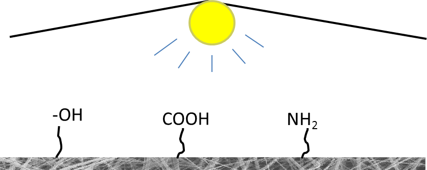
Schematic of plasma treatment of fibrous membrane to generate various functional groups.

Plasma treatment can be used to introduce oxygen-containing groups on the surface of relatively inert polymer [Wei et al 2005]. This will improve the wetting characteristic of an otherwise hydrophobic membrane therefore making it more favorable for applications this property is desirable. In cell culture, the increased hydrophilicity has been shown to facilitate cell proliferation on the membrane [Cheng et al 2013]. The activated oxygen-containing groups may then be used for grafting other molecules on the surface of the membrane. The oxygen-containing group on polysulfone (PSU) nanofiber membrane after plasma treatment was found to contain mainly of alcoholic hydroxyl group (C-OH) and this can be used to graft methacrylic acid onto it with Ce(IV) as catalyst [Ma et al 2006]. The same method has also been used to generate COOH functional groups on polycaprolactone for grafting of gelatin by using water soluble carbodiimide to active the COOH groups [Ma et al 2005]. Hoy et al (2019) used spot oxygen plasma treatment on electrospun polystyrene (ESPS) microfibers membrane to introduce specific hydrophilic zones. For their application, they used 8 layers of electrospun membrane, wet pressed together for 24hrs before plasma treatment. In their study, a 1 min O2 plasma treatment time is insufficient to fully penetrate through the membranes. A 5 min treatment rendered the membrane too hydrophilic which prevented any hydrophobic bonding between the surface and the protein to be adsorbed. A 3 min treatment time was found to be optimal for maximum adsorption of bovine serum albumin.

Despite the relative ease of using plasma treatment, a disadvantage is that the surface activation may not be uniform throughout the thickness of the membrane. Robinette et al [2005] showed that plasma radiated membrane exhibited an acrylamide concentration gradient with decreasing concentration from the exposed top surface to the relatively unexposed bottom. Kaur et al [2007] also demonstrated difference in the grafting of methacrylic acid monomer on plasma treated poly(vinylidene) fluoride membrane between the exposed surface and the unexposed bottom. Thus for thicker membrane, other stronger radiation method may be required if uniform activation across the full thickness of the membrane is required. Since plasma treatment involves modification of the surface molecule bonding, this will invariably affect its mechanical properties with a reduction in its strength and strain [Yan et al 2013]. Esmail et al (2021) conducted oxygen plasma treatment on electrospun poly(3-hydroxybutyrate) P(3HB) and copolymer poly(3-hydroxybutyrate-co-3-hydroxyvalerate) P(3HB-co-3HV) and found fractures on the fibers in particular the copolymer. P(3HB-co-3HV) fibers were also found to exhibit a slight reduction in its average fiber diameter. Such physical deterioration of the fiber is due to physical etching phenomena, where the ions in the plasma hitting the fiber surface cause erosion of the material. When medium chain length poly(hydroxyalkanoates) (mcl-PHAs) was blended with P(3HB-co-3HV) and electrospun into fibers, the resultant P(3HB-co-3HV)/mcl-PHAs fibers showed a significant reduction in fiber diameter after oxygen plasma treatment but very little fractures. Electrospun mcl-PHAs alone is unable to maintain its fiber shape due to its stickiness and tackiness at room temperature. However, when blended with P(3HB-co-3HV), this behavior of mcl-PHAs may allow the fiber to withstand fracturing during plasma treatment. Careful optimization of the treatment process is necessary to attain a balance between maximum surface treatment and retaining adequate mechancial properties.


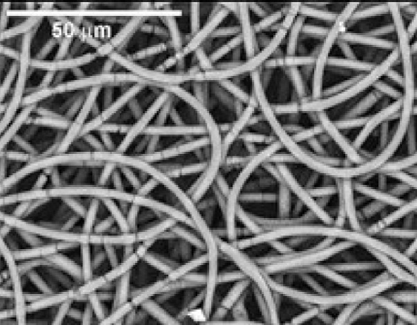
Electrospun P(3HB-co-3HV) copolymers after 12 min of oxygen plasma exposure showing fractures on the fibers [Esmail et al 2021].
Table 1. Plasma treatment condition on polymer nanofiber membrane.
Polymer (Nanofibrous membrane) Reagent / Graft material Treatment Reference
Polycaprolactone Reagent: 1-ethyl-3-(3-dimethylaminopropyl) carbodiimide hydrochloride (EDAC)

Graft material: Gelatin
Air glow discharge plasma for 5 min and electrical power of 30 W Ma Z et al 2005
Poly(L-lactic acid) Graft material: Acrylic acid monomer Oxygen plasma, gas pressure of 10-3 torr, RF power of 50 W, pulse-type negative voltage for 30 s Park et al 2006
Poly(L-lactic acid) Reagent: 1-ethyl-3-(3-dimethylaminopropyl) carbodiimide hydrochloride (EDC) and N-hydroxysuccinimide (NHS)

Graft material: Collagen and laminin
Air glow discharge plasma for 5 min and electrical power of 30 W Koh H S 2009
Polysulfone Reagent: Ce(IV)

Graft material: Methacrylic acid
Air glow discharge plasma for 5 min with gas pressure of 20 Pa and electrical power of 30 W Ma Z et al 2006
Polysulfone Graft material: Acrylamide 3 min in atmospheric pressure using a helium medium Robinette et al 2005
Poly(vinylidene) fluoride Graft material: Methacrylic acid Argon plasma treatment for 90 s with radio frequency power of 13.6 MHz and a plasma power of 30 W Kaur et al 2007

Published date: 13 November 2013
Last updated: 26 October 2021

Comments
[+]


Google Ad.