The compact nature of electrospun membrane using typical setup is the result of the fiber impacting and laying directly on top of the preceding fiber. However, if the fiber is able to sink below a liquid surface upon contact, then the on-coming fiber will be impacting the liquid surface instead of the preceding fiber. Since there is a liquid layer that separate the fibers, the fibers will be very loosely packed. Kostakova et al (2014) tested the effect of using water and ethanol for collection of polycaprolactone electrospun fibers. As expected, the hydrophobic polycaprolactone fibers float on the water surface. The fiber only starts to sink when the ethanol concentration is more than 80% in an ethanol/water mixture. Yang et al used 80% ethanol/water as collector for electrospun polyacrylonitrile (PAN) fibers. The deposited fibers were subsequently transferred to a beaker using a rod for washing followed by freeze-drying to get a fluffy 3D structure. The 3D scaffold has a pore size ranging from 15 to 20 µm as compared to electrospun PAN fibers on an aluminum foil which has a pore size of less than 5 µm. The fiber diameters from both liquid collector and aluminum foil were about 800 nm. The 3D scaffold was immersed in a chitosan solution for coating over the surface of the fibers and cross-linked to give a more stable 3D scaffold. The selection of the liquid is very important since the fiber needs to be suspended under the surface instead of floating on it. Solvents such as methanol and ethanol has been used as the liquid collector due to their low surface tension and three-dimensional structure has been constructed using this technique [Ki et al 2007, Yokoyama et al 2009, Kim et al 2014, Coburn et al 2012].
High surface area to volume ratio of the fiber meant that the fibers will either suspend just below the surface of the solvent or slowly sinking to the bottom of the solvent reservoir. Depending on the spread of the fiber deposition, a larger deposition area will result in relatively flat structure unless sufficiently long time is given for the fibers to accumulate thickness. Since the thickness is slowly built from the liquid surface, the resultant pore size may still be quite small (about 5 to 10 µm). In the study by Kim et al (2014), some steps were introduced to overcome the limitation of the basic technique. If the spinning jet can be confined to a smaller deposition area, the build-up in fiber depth will be faster. A focusing electrode in the form of a cylinder surrounding the nozzle was used for this purpose and a 3D scaffold with significant height was constructed. A continuous supply of ethanol was also introduced into the coagulation bath during fiber deposition and the resultant rising level of the coagulation bath may facilitate building a thicker scaffold. This is evident from the increased pore size with increasing ethanol feed-rate. Post-processing treatment using laser was also used to incorporate through pores in the scaffold [Kim et al 2014].
In an interesting use of this concept to include a moving collector, a rotating drum collector was used for collecting and submerging electrospun fibers through an ethanol bath followed by deposition of a next layer of electrospun fibers [Ding et al 2016]. This process continues until a sufficiently thick scaffold is constructed. Through this process, a layer of ethanol that separates the layers of electrospun fibers was introduced. This enables a rapid build up of scaffold thickness since the fibers are not tightly compacted due to the liquid layer. The low surface tension of ethanol also allows the fibers from the different layers to come into close but loose contact with one another. The rotation also facilitates alignment of the fibers. This is probably due to the lubricating effect of the ethanol on the fibers which allows movement and alignment of the fibers as the drum rotates and the flowing liquid pulls on the fibers. Such configuration has been shown to allow cell penetration and migration into the depth of scaffold.
In a study by Coburn et al (2012), the team demonstrated good cell penetration in the 3D scaffold which was constructed just by fiber deposition on ethanol bath without any external facilitation or post-spinning modifications. The material used was poly(vinyl alcohol)-methacrylate which may be more hydrophilic compared with polycaprolactone used by Kim et al (2014). A hydrophilic nanofiber may sink better in ethanol compared with hydrophobic polycaprolactone and this may result in greater pore size in the former material. Contact angle test using ethanol droplet on electrospun polycaprolactone membrane showed that it takes a couple of seconds for the ethanol droplet to fully immerse into the membrane surface [Kostakova et al 2014]. However, fiber deposition by electrospinning only takes fraction of a second and the fibers would have build up on the surface of ethanol before sinking. Instead of ethanol, Chen et al (2016) used a bath containing 7:3 (v:v) isopropyl alcohol and distilled deionized (DI) water with 0.05% Koppiphor P188 as the collector. Koppiphor P188 is a dispersing and wetting agent which facilitate submerging of the electrospun fibers as they deposit on the bath. The electrospun poly(lactic acid-co-glycolic acid) (PLGA) scaffold has a pore size distribution mainly in the 5 to 20 µm range. In vitro culture of human dermal fibroblasts showed infiltration into scaffolds up to depth of 1.4 mm after 7 days of culturing.
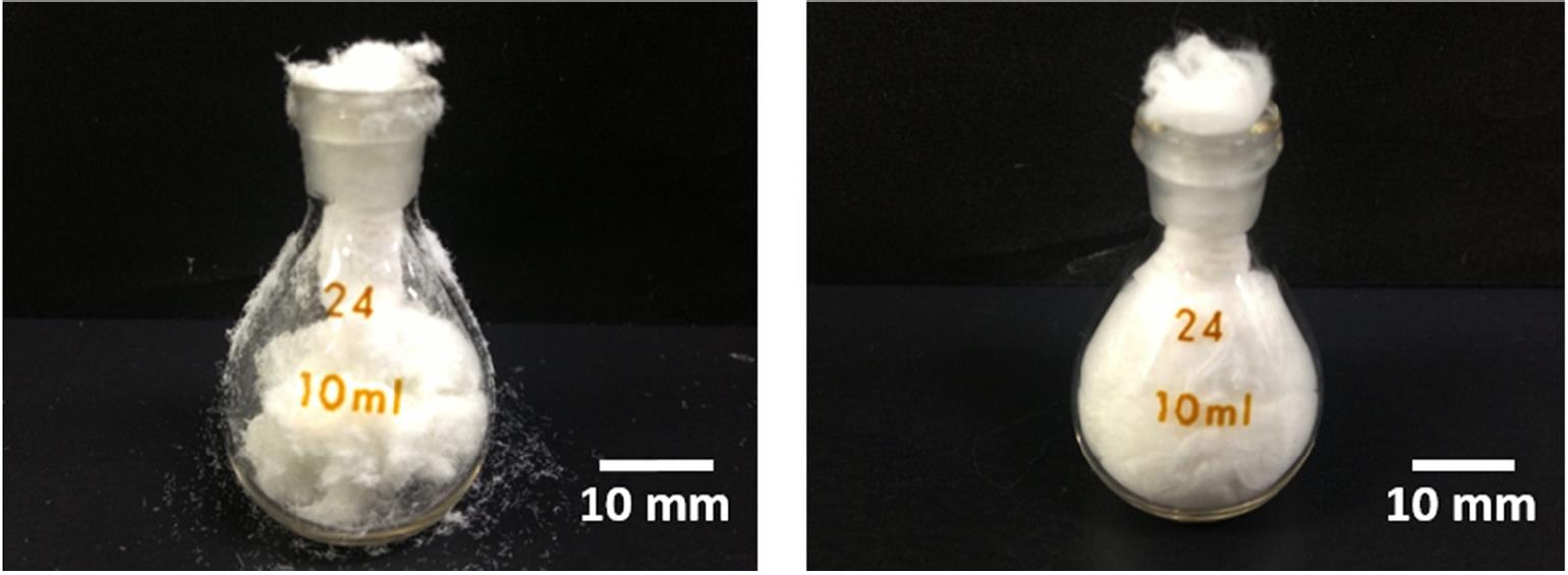
The high volume and fluffy scaffold obtained from deposition of electrospun fibers on low surface tension liquid may be subjected to compression during handling. The recovery of the scaffold following compression is dependent on its material composition. Wang et al (2015) compared the recovery of fluffy scaffold constructed from electrospun siloxane-doped vaterite and poly(L-lactic acid) (SiVPCs) fibers and core-shell fibers with SiVPCs as the core and poly(D,L-lactic-co-glycolic acid) (PLGA) as the shell material. With the presence of PLGA coating over SiVPCs fibers, the resultant scaffold was able to demonstrate recovery of about 50% while SiVPCs fibers alone showed recovery of only around 10%. Figure below illustrates the recovery potential of both scaffolds after they were squeezed into a glass bottle.
Published date: 27 August 2012
Last updated: 30 August 2022
▼ Reference
-
Chen H, Peng Y, Wu S, Tan L P. Electrospun 3D Fibrous Scaffolds for Chronic Wound Repair. Materials 2016; 9: 272.
-
Coburn J M, Gibson M, Monagle S, Patterson Z, Elisseeff J H. Bioinspired nanofibers support chondrogenesis for articular cartilage repair. PNAS 2012; 109: 100012.
Open Access
-
Ding H, Zhong J, Xu F, Song F, Yin M, Wu Y, Hu Q, Wang J. Establishment of 3D culture and induction of osteogenic differentiation of pre-osteoblasts using wet-collected aligned scaffolds. Materials Science and Engineering: C 2016; Article in Press .
-
Ki CS, Kim JW, Hyun JH, Lee KH, Hattori M, Rah DK and Park YH (2007) Electrospun Three-Dimensional Silk Fibroin Nanofibrous Scaffold. J. Appl. Polym. Sci. 106 3922-3928.
-
Kim M S, Son J G, Lee H J, Hwang H, Choi C H, Kim G H. Highly porous 3D nanofibrous scaffolds processed with an electrospinning/laser process. Current Applied Physics 2014; 14: 1
-
Kostakova E, Seps M, Pokorny P, Lukas D. Study of polycaprolactone wet electrospinning process. eXPRESS Polymer Letters 2014; 8: 554.
Open Access
-
Wang J, Zhou P, Obata A, Jones J R, Kasuga T. Preparation of Cotton-Wool-Like Poly(lactic acid)-Based Composites Consisting of Core-Shell-Type Fibers. Materials 2015; 8: 7979.
Open Access
-
Wang J, Yokoyama Y, Hattori S, Yoshikawa C, Yasuda Y, Koyama H, Takato T, Kobayashi H (2009) Novel wet electrospinning system for fabrication of spongiform nanofiber 3-dimensional fabric. Mater. Lett. 63 754-756.
-
Yang X, Chen X, Zhao J, Lv W, Wu Q, Ren H, Chen C, Sun D. Liquid-Assisted Electrospinning Three-Dimensional Polyacrylonitrile Nanofiber Crosslinked with Chitosan. Journal of Nanomaterials 2021; 2021: 4639317.
Open Access
▲ Close list