Home | About | Contact


An introduction to Adipose Tissue Regeneration

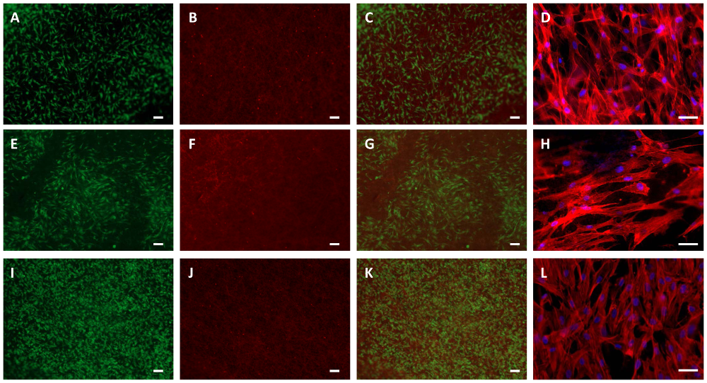
Adipose derived stem cells (ASCs) were grown on biomaterial PEUU (A-D), PEU (E-H) and PLLA (I-L) for 48 hours. Viability was detected by fluorescein diacetate (living cells; A, E, I) and ethidium-homodimer-1 (nuclei of dead cells; B, F, J) staining. C, G and K are merged pictures. Morphology was detected by TRITC-phalloidin (cytoskeleton, red) and DAPI (nuclei, blue) staining (D, H, L). Bars represent 100 µm (A-C, E-G, I-K) or 50 µm (D, H, L). Gugerell A et al. (2014) Electrospun Poly(ester-Urethane)- and Poly(ester-Urethane-Urea) Fleeces as Promising Tissue Engineering Scaffolds for Adipose-Derived Stem Cells. PLoS ONE 9(3): e90676. doi:10.1371/journal.pone.0090676.

Adipose tissue scaffold are often necessary for filling defects due to trauma, congenital malformation and oncological resection. Unlike most cosmetic and plastic surgery fillers, these defects are often of much larger volume which is a challenge for the assimilation and integration of the implant. Silicone and saline implants have been used commercially in breast augmentation. However, these permanent implants often carry the risk of rupturing and other possible long term complications. There is also no integration between the implant and the host tissue. The objective of soft tissue regeneration is to develop a scaffold that fully integrates with the host tissue or to function as a temporary scaffold until the host tissues occupy the volume defect.

To investigate the suitability of electrospun scaffolds in adipose tissue regeneration, researchers have used various materials for in vitro culturing of adipose derived stem cells (ASC). Electrospun fibers with diameters less from submicron to less than 2 µm has been shown to support ASC proliferation and differentiation. Francis et al (2012) used electrospinning to fabricate a fibrous membrane from a mixture of adipose tissue extracellular matrix and polydioxanone for culturing of adipose stem cells (ASC). The resultant scaffold was demonstrated to support ASC attachment and growth. Electrospun scaffolds made out purely of synthetic materials have also been shown to support ASC proliferation and adipogenesis. Using a mixture of Poly (L-lactide) (PLLA) and Poly (ε-caprolactone) (PCL), Chen et al (2013) was able to encourage ASC towards adipogenesis by culturing the cells in culture medium supplemented with 0.5 mM 3-isobutyl-1-methylxanthine (IBMX), 0.2 mM indomethacin 0.1 µM dexamethasone, 10 µM insulin and 1% penicillin/streptomycin. Electrospun fibrous scaffold of poly(ε-caprolactone-co-urethane-co-urea) (PEUU) and poly[(L-lactide-co-ε-caprolactone)-co-(L-lysine ethyl ester diisocyanate)-block-oligo(ethylene glycol)-urethane] (PEU) has also been demonstrated to support proliferation and differentiation of ASC to adipogenic lineage when cultured in preadipocyte differentiation medium [Gugerell et al 2014].

An important requirement for clinical utilization of adipose tissue scaffold is that it is able to fill a volume defect. While in vitro demonstration of biocompatibility and differentiation can be investigated on two-dimensional mesh, such structure is not clinically applicable and a three-dimensional (3D) scaffold needs to be constructed. Xu et al (2014) was able to electrospin fluffy 3D scaffold from soy proteins by fiber deposition on a collector that was given an opposing charge to the spinneret. Using an insulating board, 2D scaffold was constructed instead. In vitro culturing of ASC on both scaffolds showed better adipogenic differentiation on the 3D scaffold compared to 2D.

Clinical application of electrospun scaffold in soft tissue fillers may combine with current clinical procedures. Liposuction is a relatively common procedure to remove excess fats from the body. This source of fats may be used with electrospun scaffold as soft tissue fillers. A preliminary study using minced fats mixed with three-dimensional electrospun scaffold have shown that cells were still viable after 4 weeks of culture while cells in at the core of intact fat was no longer viable [Panneerselvan et al 2013]. Further, endothelial cells were also found to be viable in the tissue scaffold mixture and this may potentially accelerate the angiogenesis process.


Published date: 16 December 2014
Last updated: -

Comments
[+]


Google Ad.